Sarcoidosis is an inflammatory condition that can affect multiple organs in the body. Cardiac sarcoidosis is a rare type in which clusters of white blood cells, called granulomas, form in the tissue of the heart. Affecting about 5% of people who suffer from sarcoidosis, it can impact the electrical system of the heart and may lead to arrhythmias or heart failure.
Three Ottawa Heart Institute presentations at the 2016 Canadian Cardiovascular Congress provided new information on the role of imaging technology in diagnosing and understanding cardiac sarcoidosis.
One study, by resident Daniel Belliveau, MD, and colleagues looked at whether an imaging phenomenon called late gadolinium enhancement (LGE) on cardiac magnetic resonance imaging (MRI) could help to predict risk of adverse cardiac events in patients with sarcoidosis. LGE can identify different types of cells within the heart, such as scar tissue or granulomas. The researchers looked at data from 13 previously published studies of more than 1,300 patients with sarcoidosis. Patients who did not have LGE on their MRI scans had a decreased risk of major adverse cardiac events and death. Patients who did have LGE (about 25%) had an increased risk.
Cardiology resident Juan Russo, MD, presented results of a study looking at whether chest X-ray, chest CT or MRI best identified cardiac sarcoidosis. He and his colleagues used a database of patients with known sarcoidosis who had undergone chest imaging. They read all images without knowing whether the patients had eventually been diagnosed with cardiac sarcoidosis as well and identified 51 patients who had one or more imaging scan that appeared to show the disease.
Of these, 22 patients actually had cardiac sarcoidosis. The ability to detect the condition was lowest for chest X-ray at 30%, compared to 86% for MRI and 90% for chest CT. However, more patients had received chest X-ray than chest CT or MRI. “Chest X-ray appears to be suboptimal as a screening test for patients in whom cardiac sarcoidosis is suspected,” wrote the researchers.
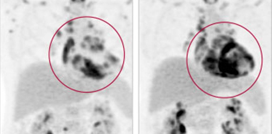
Fellow Daniel Juneau, MD, and his colleagues in the Cardiac PET Research Team examined patterns of sarcoidosis outside of the heart in patients who underwent a type of imaging called positron emission tomography (PET)-CT after being diagnosed with acute cardiac sarcoidosis. In the 18 patients studied, almost 90% also had disease in their lymph nodes and about 60% had disease in organs other than the heart. The percentage that had sarcoidosis in the lungs (just under 40%) was much lower than in previous studies. Only one patient had disease confined to the heart, known as isolated cardiac sarcoidosis.